


















Electromyography (EMG)
Content
Description
Electromyography (electromyelography) - EMG is an examination of nerves and muscles, and we distinguish 2 types of examination:
- 1. Stimulation EMG (nerve conduction examination)
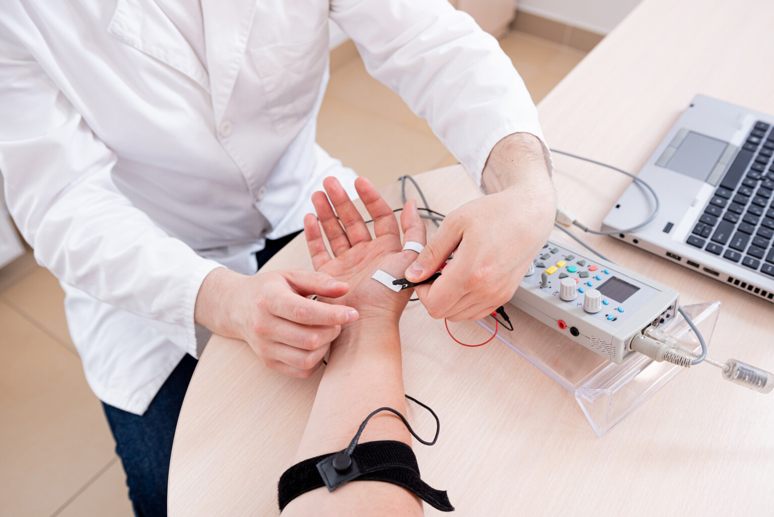
An examination in which electrodes are placed on the skin, which send a weak electric current. The patient feels the muscle twitch as a gentle pinch.
This examination measures how quickly and how well the nerve conducts impulses.
- 2. Needle EMG (used to examine muscles)
A thin electrode (fine needle) is inserted into the muscle and after sending a weak current, the muscle activity is measured at rest and in movement.
Facts
- Outpatient procedure
- Examination lasts 20 - 40 min
EMG is used to diagnose muscle and nerve diseases, for example:
- Carpal tunnel syndrome (canal)
- Radiculopathy - nerve compression when a disc compresses them
- Polyneuropathy (and its causes - e.g. diabetes, alcoholic neuropathy)
- Muscle diseases (stiffness, muscle pain, myopathies), neuromuscular transmission diseases (myasthenia gravis) and motor nerve disorders (ALS - amyotrophic lateral sclerosis)
- Nerve damage after an injury
- Examination of the causes of tingling in the limbs, cramps, tics, tetany
The examination takes place in an outpatient clinic and does not require any special preparation - it is recommended that the skin is not too oily (therefore, do not apply cream or oil).
In some cases, an EMG examination is not recommended because it may pose a risk to the patient. Therefore, inform your doctor if:
- you have a pacemaker
- you are taking blood thinners
In such cases, a neurologist will consider whether the examination can be performed.