


















Dental X-rays
Content
Description
Teeth are hard structures of enamel and dentin that are very good visible on X-rays (they are X-ray contrast). The teeth are fixed in the jawbone. X-rays can also show hidden decay (caries) in the spaces between the teeth, below the level of the gums or those under fillings or dental crowns. It also detects foreign bodies, congenital disorders and injuries.
Modern X-ray devices generate a minimal amount of radiation and capture the image digitally, so that the image can be edited afterwards. In this way, the images provide detailed information about the teeth, the bones and the surrounding tissues.
X-ray is used during preventive examinations (detection of tooth decay) and before dental procedures (for example when planning dental braces, dental implants, crowns, bridges, teeth extraction and in dental prosthetics).
Facts
- Painless procedure
- Outpatient procedure
- Image is ready within 10 minutes
- No restrictions before examination
- X-ray of the tooth (intraoral image) - X-ray of individual tooth, which evaluates the detail of the tooth, the depth of tooth decay, the condition of the roots and the amount of tartar.
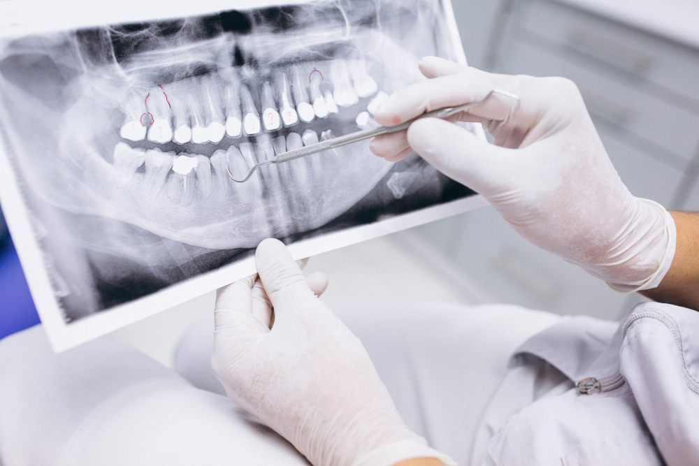
- Panoramatic X-ray (OPG, orthopantomography, orthopantomogram – is a panoramic X-ray that shows the entire dentition. On this image, the entire bite can be seen and the relative position of the teeth and the surrounding structures can be revealed.
- CT (Computed Tomography) - serves for 3D imaging. Necessary before planned major interventions for evaluation of tooth decay, inflammation, cysts, tumors, diseases of bone tissue and the temporomandibular joint, complications after injuries. Modern devices rotate around the standing patient.
Patients wear a lead vest to protect against radiation, which prevents the penetration of X-rays.
X-rays are not performed on pregnant women in the first trimester and small children.
In such cases, only necessary images are taken when the life of the mother or child is at risk.

Related procedures
 Dentistry
- Dental Implants
- Teeth whitening (Bleaching)
- Preventive dental care
- Dental hygiene
- Tooth decay (Dental caries treatment)
- Dental pulp and dental root treatment (Endodontics)
- Dental braces (Orthodontic cases)
- Dentures (False teeth)
- Dental crowns and Dental bridges and Veneers
- Tooth extraction
- Dental Treatment Under General Anesthesia
- Periodontitis (periodontal disease)